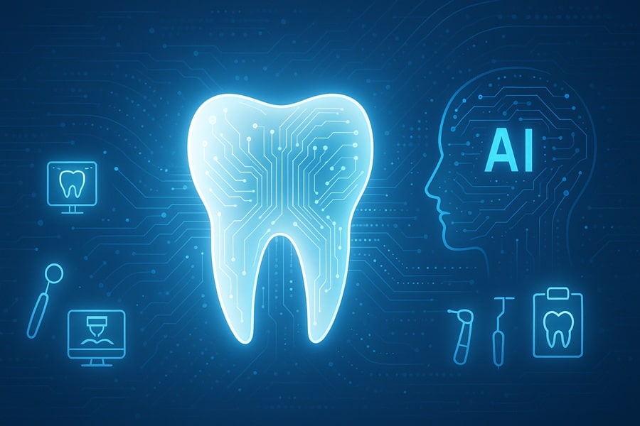
AI and digital dentistry in 2026: From Automated Image Interpretation to Treatment Design and End to End Clinical Workflow Integration

This article was medically reviewed by Diane Boval, DDS, a licensed dentist practicing in California.
AI and digital dentistry touches almost every part of care in 2025—from reading images to designing restorations and orchestrating the entire clinical workflow. If you’re a dentist in Southern California or searching for a dentist near me, this guide explains what’s real today, what’s coming next, and how teams can adopt these tools safely and ethically. Whether you’re scheduling routine dental checkups or planning a smile makeover, understanding digital tools helps you make informed choices.
This guide starts with the basics—what the technology covers—and then moves through specific applications such as automated image interpretation, computer‑aided design and manufacturing, predictive analytics, robotics, teledentistry, and virtual reality. Each section explains how the tools work, who benefits, and the precautions needed to protect patient privacy and clinical judgement. Throughout, we use clear headings, short paragraphs, and numbered lists so you can quickly find the information most relevant to you.
What Is AI and digital dentistry Today?
AI and digital dentistry blends imaging AI, computer‑aided design and manufacturing, 3D printing, robotics, and cloud software to support diagnosis, treatment planning, fabrication, and documentation within one workflow. Modern systems use machine learning algorithms trained on diverse datasets of radiographs, cone beam CT scans, intraoral scans, and clinical records to detect patterns that signal disease or inform treatment.
In digital radiography, neural networks identify caries, bone loss, periapical lesions, and nerve proximity. In CAD/CAM, algorithms suggest margin lines, occlusal schemes, and aligner stages. Predictive analytics combine imaging with risk factors—such as smoking or occlusal forces—to forecast fracture risk or peri‑implant disease. Robotics assist with drilling and implant placement, while teledentistry platforms use AI triage to prioritize appointments. Together, these tools transform how care is delivered and documented.
To work well across different clinics, training datasets must include pediatric and adult cases, mixed dentitions, orthodontic appliances, and restorations. Otherwise, models may perform poorly when the clinic switches sensors or when exposure settings change. Versioned releases and clear “what changed” notes help clinicians understand why a model behaves differently after an update.
These datasets also need to represent all age groups. For example, integrating data from pediatric dentistry ensures that growth patterns and developing dentitions are considered, while cases from older adults help the models recognize restorations and bone density changes. Without this balance, AI tools may struggle when applied to new demographics.
Imaging AI: Automated Image Interpretation from Radiographs to CBCT
Computer vision models now read bitewings, periapicals, panoramic radiographs, and CBCT volumes to flag cavities, periapical changes, bone loss, and anatomical risks. Sensitivity and specificity vary by lesion depth and device type but generally range around 80–92 % and 75–90 %, respectively (Luke et al., 2025). Early enamel lesions remain harder to detect than dentin‑level caries, but contrast normalization and multi‑view fusion improve performance. These systems also help detect early periodontal changes—such as bone resorption and pocketing—that may signal the need for gum disease therapy.
On CBCT, convolutional neural networks map nerve canals, sinus cavities, and root morphology. Distance errors typically range from 0.5–1.2 mm for nerve proximity identification, which aids guide design but still requires clinical confirmation. Quality control dashboards report failed segmentations and out‑of‑range exposures so tech leads can recalibrate sensors or update presets after firmware changes.
Clinicians remain at the center: AI provides overlays and measurements but doesn’t replace clinical judgement. If a system flags a lesion, the provider verifies its presence and documents an override if the result conflicts with clinical signs. These human‑in‑the‑loop checks maintain safety and accountability.
Intraoral Scanning, CAD/CAM, and 3D Printing
Digital impressions capture the dentition with high precision, feeding CAD/CAM software and additive manufacturing systems. Accuracy depends on calibration routines, pass strategy, and stitching algorithms. Weekly checks with calibration panels and a five‑minute tolerance test keep scanners aligned. For CAD/CAM, margin smoothing and cusp reinforcement balance esthetics with strength; validated libraries match local materials such as zirconia, lithium disilicate, or hybrid resin ceramics. Chairside milling and printing mean that crowns and bridges can often be designed and fabricated in a single visit, reducing the number of appointments and temporary restorations.
3D printing allows chairside provisionals and surgical guides. Resin selection, post‑cure protocols, and nesting orientation affect strength and margin accuracy. Studies show that using validated printer profiles and layer heights reduces remake rates by 10–25 % (Migliorati et al., 2025). For surgical guides, metal sleeves, window positions, and offsets are tested on a bench model before patient use. Color and translucency benefit from photo alignment—using neutral reference cards and a fixed light source—to ensure shade consistency across visits and locations.
Machine Learning and Deep Learning Models
Most dental AI tools rely on supervised learning algorithms such as convolutional neural networks, U‑Net architectures for segmentation, and transformer‑based models for sequence prediction. These networks learn from thousands of annotated images, identifying patterns that human eyes may miss. For example, segmentation networks can delineate enamel, dentin, pulp, periodontal ligament, and alveolar bone on CBCT slices, providing measurements for bone height and thickness. Landmark detection models identify apices, canals, and cephalometric points for orthodontic analysis.
Training quality matters. Datasets must be balanced across ages, ethnicities, and device types to avoid bias. Models should also be evaluated on external test sets rather than just the training clinic’s data. Many vendors now provide performance metrics—including mean absolute error for bone measurements and confusion matrices for lesion detection—so clinicians can compare tools. Emerging generative models can simulate missing slices or create synthetic training data, potentially improving robustness.
Predictive Analytics and Personalized Dentistry
Beyond detection, AI combines imaging data with patient habits to forecast risk and personalize care. For example, predictive models integrate plaque scores, smoking status, diet, and salivary flow to estimate caries risk. In periodontology, combined indices that include plaque, bleeding on probing, and bone level changes drive tailored recall intervals and home‑care scripts. For restorative cases, fracture risk models consider occlusal scheme, parafunction, and remaining wall thickness to suggest material choices or staging.
In orthodontics, adherence models flag patients who may struggle with aligners or nightguards. The system can generate reminders and short check‑in notes, using plain language and pictures to encourage compliance. These nudges reduce non‑tracking events and improve long‑term outcomes. Predictive analytics also support preventive counseling by highlighting modifiable risk factors and quantifying potential benefits of interventions.
AI‑Enhanced Orthodontics and Prosthodontics: Planning and Design
Automated staging tools suggest aligner steps, bracket positions, and elastic patterns while showing expected root torque and anchorage loads. Fixed protocols draw from libraries of bracket prescriptions that reduce manual edits and keep consistency across providers. Deep learning models generate margin lines, connector sizes, and occlusal schemes for crowns and bridges, matching chewing patterns and esthetic zones. Clinical studies in 2025 report time savings of 30–45 minutes per case and remakes reduced by about 18 percent when AI‑assisted CAD tools are used (Migliorati et al., 2025).
Predictive checks evaluate interproximal contacts, path of insertion, and minimal thickness. When a bridge or hybrid prosthesis is designed, connectors are tested against expected chewing forces; the system flags unsupported spans. For aligner cases, non‑tracking risk is flagged early using features such as rotation angle, root length, and periodontal support. All plans, measurements, and snapshots are saved with version numbers so changes are traceable. Labs receive structured tickets with materials, shade, try‑in steps, and delivery checks.
Robotics and Surgical Assistance in Oral Surgery
Robotic guidance systems assist clinicians during implant placement and complex extractions. These platforms use pre‑operative CBCT data to plan drill trajectories and then guide the handpiece to the correct position and depth. A semi‑active robotic arm maintains orientation while allowing the dentist to control velocity and tactile feedback. Early clinical trials report improved accuracy in implant positioning, with deviations less than 1 mm, and reduced surgery time by about 15–20 percent. Despite these advances, thoughtful anaesthesia and anxiety control remain essential—nervous patients may benefit from sedation dentistry so they remain calm and comfortable throughout the procedure.
Robots also standardize repetitive tasks such as polishing and cleaning implant surfaces during surgery. For impacted third molars, robotic arms integrated with haptic feedback help residents learn proper angulation and force application. As these tools develop, regulatory bodies require proof of safety, fail‑safe modes, and clear guidelines on human oversight.
Because robotic guidance is most frequently applied in complex extractions and implant placements, these innovations are deeply connected with oral surgery and require specialized training. Dentists should still counsel patients on potential risks and discuss traditional surgical options when appropriate.
Teledentistry and Virtual Consultations
AI extends beyond brick‑and‑mortar clinics. Teledentistry platforms use chatbots and image triage to screen patients remotely. For example, a patient uploads a smartphone photo of a lesion; the system classifies its urgency and suggests a follow‑up within 24 hours, a routine visit, or immediate referral. This approach is particularly useful for triaging urgent cases—such as trauma, swelling, or severe pain—that may require immediate emergency dentistry rather than waiting for a scheduled appointment. Automated triage improves access and ensures that high‑risk cases are prioritized when schedules are tight.
Remote monitoring also supports clear aligner therapy. Patients scan their teeth using an at‑home device; AI detects fit issues and schedules an intervention only when needed. For post‑operative care, virtual check‑ins reduce unnecessary trips and alert providers to complications early. Emerging platforms integrate into the electronic health record so notes, images, and messages are stored in one place.
Virtual and Augmented Reality in Dental Education and Patient Care
Virtual reality (VR) and augmented reality (AR) enhance both training and patient experiences. Dental students can practice procedures in a VR simulator with real‑time feedback on depth, angle, and force, building muscle memory before treating patients. AR overlays onto the operative field display nerve pathways, root morphology, or drill depths during surgery, improving situational awareness.
For patients, VR headsets provide immersive distraction during lengthy procedures, reducing anxiety and perceived pain. AR apps allow them to visualize the planned outcome—such as the appearance of veneers, orthodontic correction, or even brighter smiles from teeth whitening—before treatment begins. These technologies rely on accurate 3D models generated from intraoral scans and CBCT data, highlighting the interplay between imaging and immersive media.
End‑to‑End Clinical Workflow Integration and Automation
AI and digital dentistry tie capture software, planning tools, fabrication platforms, and the electronic health record together so findings, plans, and consents live in one place. Integration reduces redundant data entry: orders flow from planning to the lab ticket, and images with overlays attach to the chart automatically. Version history captures who edited margins or implant positions, when, and why. This traceability is essential for teaching clinics and multi‑location groups.
Hand‑offs are defined: the imaging technician submits the study, a reviewer signs findings, and the planner confirms that guides and models match the signed version. Clear role definitions—who reviews, who signs, who edits—keep accountability strong and reduce emergency remakes. Connected platforms also facilitate scheduling, consent management, and billing, turning a series of isolated tasks into a coordinated workflow.
Privacy, Security, and Ethical Considerations
De‑identified datasets, role‑based access, and audit trails protect patient privacy. Consent forms must explain how data may train or improve tools and provide opt‑out paths. HIPAA and GDPR rules require encryption, strict access control, and logging of every change. Bias testing across device types, ages, and dentitions reduces unfair errors; training sets should reflect the diversity of real‑world patients.
Ethical questions go beyond privacy: How do we ensure that AI does not widen disparities? What happens when a model misclassifies a rare pathology? Clear policies state that outputs are advisory and that licensed providers make final decisions. If an AI suggestion conflicts with clinical signs, the provider documents the rationale for override. Clinics should maintain review sets to check model performance after each software update and report issues to vendors for correction.
Change Management: Training, SOPs, and Adoption
Successful adoption begins with a single high‑value use case, a short standard operating procedure (SOP), and regular feedback huddles. Track three metrics: accuracy gaps versus reviewers, time saved, and unintended effects (like increased retakes). Train two “super‑users” who maintain presets, retrain staff, and liaise with vendors for updates. Weekly five‑minute huddles allow team members to raise friction points and celebrate wins.
Set a measurable goal for the first 90 days—fewer retakes, fewer remakes, or faster planning—and post it where everyone can see. Celebrate progress and adjust protocols as needed. Quarterly meetings with vendors ensure that software updates align with clinical needs and that new features remove clicks rather than add complexity.
Costs, ROI, and Insurance Considerations
Licenses may be per operatory, per provider, or per study. Pricing models range from low‑volume per‑study plans to site licenses for busy clinics. Contracts should specify data export formats, model versioning plans, uptime guarantees, and support response times. Ask vendors for validation data on the exact devices you own and for calibration guidance.
Return on investment shows up as fewer remakes, shorter seat times, and better patient acceptance due to clearer visuals. Even small time savings compound across a year in a multi‑operatory clinic. Insurance considerations include billing codes for AI‑assisted diagnostic interpretations and whether carriers reimburse for digital impressions or 3D‑printed provisionals. Practices should track these costs and savings to inform future purchases.
Regulations, Standards, and Governance of AI in Dentistry
Regulatory frameworks have matured. In the United States, many AI dental imaging platforms are classified as Class II medical devices, requiring transparency in training datasets and post‑market monitoring. The European Union’s AI Act demands algorithmic explainability and bias audits. HIPAA updates in 2025 reinforce encryption, access logs, and explicit consent. Dental organizations such as the ADA, FDI, and ISO TC106 are developing validation benchmarks for AI in radiography, orthodontic simulation, and restorative design.
The future will rely on “AI passports”—digital records of model lineage, accuracy, and retraining frequency—to ensure clinical safety and accountability. Dental schools now teach students how to interpret performance metrics, verify dataset diversity, and understand legal requirements. Understanding these standards is an essential skill for modern practitioners and researchers.
The Road Ahead: Future Trends and Conclusion
AI and digital dentistry will continue to evolve. Generative models may design restorations from natural‑language descriptions, and reinforcement learning could optimize orthodontic treatment sequences. Integration with electronic health records will improve, enabling automatic risk alerts and personalized education materials. Robotics will become more autonomous but will still require human supervision. Virtual and augmented reality will be more immersive and realistic, and teledentistry will expand access in underserved regions.
If you’re a dentist in Brea, CA or exploring options with a dentist near me, embracing these technologies can improve consistency, efficiency, and patient satisfaction. Gold Coast Dental’s team is ready to guide you through adoption, from selecting the right tools to training staff and integrating them into your workflow. Contact us today to schedule a consultation or visit one of our Southern California offices.
FAQ
How does AI improve diagnostic accuracy?
AI standardizes detection and highlights subtle findings across images. Clinicians confirm and prioritize care, and override suggestions when they conflict with clinical judgment.
Will AI replace dentists?
No. AI is a support tool. Final diagnosis, treatment planning, and hands‑on procedures remain clinical decisions made by licensed providers.
Which images benefit most from AI?
Bitewings and periapicals help detect caries and bone loss; CBCT aids in mapping anatomy and planning surgical guides.
What about privacy?
Systems must follow HIPAA and GDPR rules, with strict access control, encryption, and opt‑out provisions in consent forms.
Does AI change the visit length?
After a learning curve, most clinics see time savings in planning and documentation. Longer appointments may be needed for training and calibration.
Is 3D printing reliable for final restorations?
Yes, when the printer, resin, and curing profile are validated and monitored. Regular calibration and protocol adherence are key.
How do we validate a model?
Compare outputs to local ground truth on a sample set, monitor drift after software updates, and keep a small review set for ongoing checks.
What training does staff need?
Short SOPs, hands‑on sessions, and quick refreshers whenever software updates introduce new features or workflows.
Refrences :
Last reviewed November 2025.

